
The numbers, honestly数字,老实说
What the simulation says, including where it fails模拟说了什么,包括没做好的地方
Every result here comes from synthetic gels with a known truth. Synthetic data tests the analysis pipeline against our own assumptions, not the chemistry: a real build would have to repeat every check on real gels.
这里的每个结果都来自“已知标准答案”的合成凝胶。合成数据只能拿我们自己的假设去检验分析流程,检验不了化学;真造出来以后,每一项检查都要在真凝胶上重做一遍。
Read this first. This section is version 1, measured on gels from our own generator. The only test built by a separate AI agent's generator, red-team gel S3 (see Tested harder), went worse: per-myofibril axial error 1.8% RMS with 7.5% beyond ±2%, a transverse bias of −17.8%, and 0% of the under-expanded patch flagged. Treat everything below as development results.
先看这里。这一节是第一版,测的是我们自己的生成器造的凝胶。唯一一块由另一个 AI 智能体的生成器造的考试凝胶 S3(见更严的考试)结果更差:每根肌原纤维轴向误差均方根 1.8%,7.5% 超过 ±2%;横向偏差 −17.8%;膨胀不足的区域只标出了 0%。下面的一切都请当作开发阶段的结果来看。
The first estimate was too optimistic, and the simulation said so第一次估计太乐观了,是模拟自己揭穿的
simulated模拟
The first desk calculation said one myofibril segment of 10 sarcomeres gives its expansion to 0.21–0.35% (RMS), for assumed before-image contrast of 5–50%. It assumed perfectly regular sarcomeres. Real ones vary by about 3% from one to the next, and under that jitter (and at some sarcomere lengths) the original estimator (a free-frequency fit) locked onto side-lobes: 10–17% of myofibrils came out wrong by 7–19%. The first full whole-gel simulation failed 15 checks.
第一次纸面计算说:一段含 10 个肌节的肌原纤维,放大倍数能测准到 0.21–0.35%(均方根误差),前提是膨胀前图像的对比度在 5–50% 之间。可这个计算假设肌节长得完全一样。真实肌节一节和一节之间差约 3%,原来的估计方法(自由频率拟合)一遇到这种抖动(以及某些肌节长度)就会锁到旁瓣上:10–17% 的肌原纤维被算错 7–19%。第一次整块凝胶模拟因此挂掉了 15 项检查。
The fix is the Z-disc ladder: the Z-discs in the expanded image define a sarcomere-by-sarcomere coordinate, and the before-image is fitted on that ladder, so the jitter is carried along instead of fought. Segments need at least 8 sarcomeres. Result: 0.3–0.6% RMS per myofibril across simulation runs, 0.32–0.48% across the four gels of the final run and 0.35% on the blind gel, with at most 0.48% of myofibrils off by more than ±2%.
修正办法叫 Z 盘梯子:用膨胀后图像里的 Z 盘标出一节一节的肌节坐标,再沿着这把“梯子”去拟合膨胀前的图像,抖动被顺着带走,而不是硬扛。每段至少要 8 个肌节。结果:各轮模拟中每根肌原纤维的误差为 0.3–0.6%(均方根),最终一轮四块凝胶为 0.32–0.48%,盲测凝胶为 0.35%;偏差超过 ±2% 的肌原纤维最多占 0.48%。
The error bar went up, and the simulation itself raised it.
误差范围变大了,而且是模拟自己调大的。
Our own blind gel (version 1, development)我们自己的盲测凝胶(第一版,开发用)
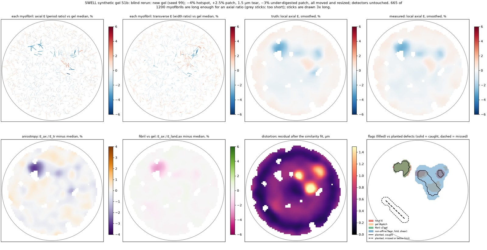
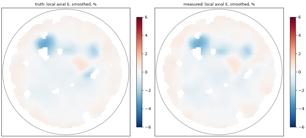
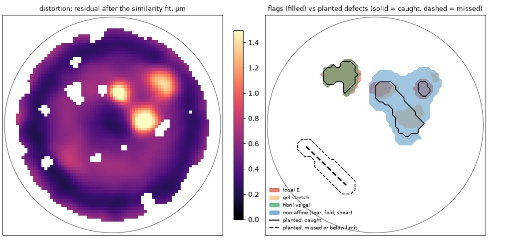
Gel S1b was our own blind test: the same generator with a new random seed, every planted defect moved and resized, and nothing tuned after it ran. It is not independent, because our generator shares our assumptions; red-team gel S3 is the red-team test. It holds 1,200 myofibrils in a 1.5 mm punch expanded 4.5×.
S1b 号凝胶是我们自己的盲测:同一个生成器换了随机种子,所有埋进去的缺陷都换了位置、改了大小,跑完以后什么参数都没再调。它不算独立,因为生成器和我们有同样的假设;红队检验是凝胶 S3。它在一块 1.5 mm 的胶片里放了 1,200 根肌原纤维,放大 4.5×。
| Planted defect (blind gel)埋入的缺陷(盲测凝胶) | Size大小 | Flagged by its own detector被对应探测器标出 | Flagged by any detector被任一探测器标出 | Result结果 |
|---|---|---|---|---|
| Under-expanded hotspot局部膨胀不足 | −4% | 100% | 100% | PASS通过 caught抓到 |
| Under-digested patch消化不足区 | −3% | 100% | 100% | PASS通过 caught抓到 |
| Stretched patch被拉伸的区域 | +2.5% | 51% | 100% | FAIL失败 flagged everywhere, but by the non-affine detector: right place, wrong label整片都被标出了,但标它的是“非仿射”探测器:位置对,类别错 |
| Tear裂缝 | 1.5 µm | never crosses the detector threshold信号始终没超过探测阈值 | BELOW LIMIT低于下限 missed; the limit is ~2 µm漏掉;探测下限 ~2 µm | |
Pass rule: the detector named for the defect must flag at least 80% of its area.
合格标准:该缺陷对应的探测器要标出至少 80% 的面积。
The two misses, plainly: a mild +2.5% stretch is caught under the wrong label, and a 1.5 µm tear is too small to see. Across all four synthetic gels, 31 of 33 checks pass, 1 fails and 1 is below the detection limit. Three of the gels (S0, S1, S2) are development gels, because the detectors were fixed while looking at S1; S1b was our own blind gel, and S3 has since replaced it as the red-team test. On development gel S1, a 2 µm tear was flagged over 89% of its area.
两处没做好,直说:轻微的 +2.5% 拉伸被标错了类别,1.5 µm 的裂缝太小看不出来。四块合成凝胶合计,33 项检查中 31 项通过,1 项失败,1 项低于检测下限。其中 S0、S1、S2 三块是“开发用”凝胶:探测器是对着 S1 调好的;S1b 是我们自己的盲测凝胶,后来由 S3 取代成为红队检验。在开发凝胶 S1 上,一条 2 µm 的裂缝被标出了 89% 的面积。
Coverage: about half the myofibrils carry the axial ruler覆盖率:约一半肌原纤维能用轴向尺子
Every myofibril gets the transverse (width) ruler and the landmark (position) ruler. Only segments of at least 8 sarcomeres get the axial period ratio: 665 of 1,200 (55%) in the blind gel and 626 of 1,200 (52%) in the development gels. In real meat this would depend on how gently the tissue is homogenised.
每根肌原纤维都能用横向(宽度)尺子和地标(位置)尺子。但只有至少 8 个肌节长的片段才能算轴向周期比:盲测凝胶的 1,200 根里有 665 根(55%),开发凝胶为 626 根(52%)。换成真肉,这取决于打碎组织时有多温柔。
Transverse isotropy and the shape-aware ruler横向各向同性与“认形状”的尺子
A gel can swell evenly along a myofibril and unevenly across it. So the pass criteria include transverse isotropy, Eax ÷ Etr − 1 within ±3% (95% CI, n ≥ 200), and the axial and transverse factors are always reported separately. On the blind gel the transverse factor per myofibril is 0.00% ± 0.50% (bias ± SD).
凝胶可能顺着肌原纤维胀得很匀,横着却不匀。所以合格标准里加了横向各向同性:Eax ÷ Etr − 1 落在 ±3% 以内(95% 置信区间,n ≥ 200),轴向和横向的放大倍数永远分开报告。盲测凝胶上,每根肌原纤维的横向放大倍数偏差为 0.00% ± 0.50%(偏差 ± 标准差)。
The width ruler is the fragile one: it needs a model of each cross-section. A boxy cross-section fitted as an ellipse is off by −9.0% to −15.2%, depending on the before-image method. So the pipeline measures each myofibril's cross-section shape from the expanded z-stack, and the bias drops to ≤ 0.04%.
宽度尺子最娇气:它得知道每根纤维的横截面长什么样。把方形截面当椭圆来拟合,会差 −9.0% 到 −15.2%(取决于膨胀前图像的拍法)。所以流程改成从膨胀后的 z 叠层里直接量每根纤维的截面形状,偏差降到 ≤ 0.04%。
Blur calibration still matters. If the before-image blur is calibrated 5% too wide, the bias is 1.05–3.23%, so the first gate calibrates it to ±2%. At that target the worst bias in our own model is 1.09%. Red-team gel S3 showed the model is not enough: a blur shape the ruler does not model biased it by −17.8%, with the right blur widths or not.
模糊标定照样要紧。膨胀前图像的模糊如果标宽了 5%,偏差就是 1.05–3.23%;所以第一道关卡要求标定到 ±2%。在这个精度下,按我们自己的模型最坏偏差是 1.09%。红队凝胶 S3 证明这个模型不够:一种标尺没建模的模糊形状让它偏了 −17.8%,模糊宽度给没给对都一样。
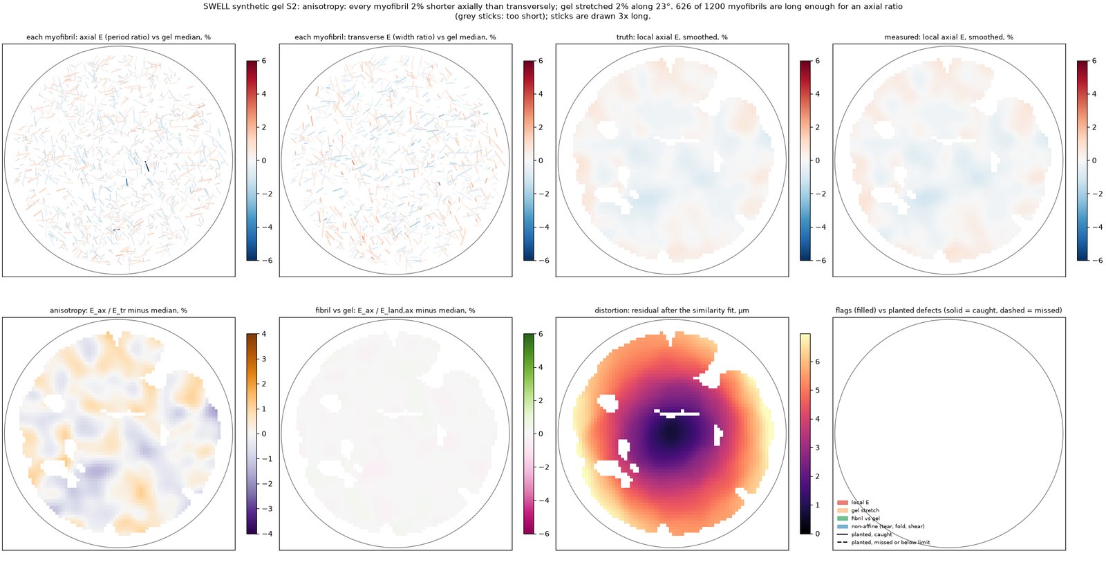
On development gel S2, a planted 2% anisotropy is recovered as −2.12% [−2.37%, −1.87%] (truth −2.03%), and a planted gel stretch of 2.00% along 23° is found by the orientation test as 2.03% along 23°.
在开发凝胶 S2 上,埋入的 2% 各向异性被测成 −2.12% [−2.37%, −1.87%](真值 −2.03%);埋入的整体拉伸(沿 23° 方向 2.00%)被方向检验测成沿 23° 方向 2.03%。
Development gel S2: anisotropy and gel stretch开发凝胶 S2:各向异性与整体拉伸
Optics, on paper光学,纸面结果
simulated模拟
Sampling: 0.194 µm pixels against a Nyquist pixel of 0.200 µm, i.e. 1.03× Nyquist; the 10× track gets 0.84×.
Focusing into water: a dry objective imaging into a water gel suffers spherical aberration, but the Gibson–Lanni model gives the 4× objective a Strehl ratio, relative to the gel's surface, of at least 0.98 over 0–12 µm (index mismatch only: it leaves out the objective's own aberrations, its tube length and any coverslip error); worst over the specimen depth 0.99 (4×) and 1.00 (10×). Lateral PSF width 0.412 µm.
采样:像素 0.194 µm,奈奎斯特要求 0.200 µm,即奈奎斯特的 1.03×;10× 路线为 0.84×。
对焦进水里:干物镜看进水凝胶会有球差,但 Gibson–Lanni 模型算出 4× 物镜在 0–12 µm 深度内、相对凝胶表面的斯特列尔比不低于 0.98(只算折射率失配:没算物镜自身的像差、筒长和盖玻片误差);在样本厚度范围内最差为 0.99(4×)和 1.00(10×)。横向点扩散函数宽 0.412 µm。
simulated模拟
One gel at 4.5×: 804 tiles per plane × 10 planes, 25.5 GB of raw data, 1.3–2.2 h of scanning.
一块放大 4.5× 的凝胶:每层 804 个视野 × 10 层,原始数据 25.5 GB,扫描 1.3–2.2 h。
Model limits: magnification comes from a thin-lens model (a ray-traced model was planned but not done), and the camera's noise figures are assumptions not yet checked against a datasheet.
模型的局限:放大倍数用的是薄透镜模型(原计划换成光线追迹模型,但没有做),相机噪声参数是假设值,还没对照数据表核实。
open problem未解决
Not enough light in the worst case最坏情况下光不够
Widefield optics see the whole thickness of an expanded myofibril, so out-of-focus light adds a background pedestal. At the lowest labelling simulated (0.5% of amines) on a 2 µm myofibril, the signal-to-noise ratio falls 12.4 → 5.6 against a target of 10. Reaching the target needs 3.2× the dose (exposure × irradiance), which costs bleaching. Before the pedestal was modelled, the first pass had given 12–42 across the labelling range.
宽场光学会把膨胀后肌原纤维的整个厚度都收进来,焦外的光叠成一层背景“底座”。在模拟的最低标记率(0.5% 的氨基被标记)、2 µm 粗的肌原纤维上,算上这层底座后,信噪比的变化是 12.4 → 5.6,而目标是 10。要达标得把光剂量(曝光时间 × 光强)加到 3.2×,代价是荧光漂白。没算这层底座之前,第一轮估计在不同标记率下为 12–42。
Not solved. The named routes were a dose budget, deconvolution and a dye titration.
没有解决。已经点名的出路有三条:光剂量预算、反卷积、染料浓度梯度试验。
Tested harder更严的考试
A fresh AI review found the weak spots. Here is what changed.一次全新的 AI 审查找出了薄弱处。下面是改了什么。
On 9 October 2026 a fresh AI review session (another Claude instance that had not built anything here, not a person) reran the download: the fingerprint matched, all 11 tests passed and every number reproduced. Then it listed what the tests could not show. Each point below is fixed, or stated as an open problem with its numbers. The first version was withdrawn on 10 October 2026, because it carried the original brief's superseded home-lab plan.
2026 年 10 月 9 日,一个全新的 AI 审查会话(另一个没参与过这里任何构建的 Claude 实例,不是人)把下载包重跑了一遍:指纹对得上,11 个测试全部通过,每个数字都能复现。然后它列出了这些测试证明不了的地方。下面每一条,要么已经修好,要么作为一个带数字的未解问题写明。第一版已于 2026 年 10 月 10 日撤回,因为里面还带着原始任务书里已作废的居家实验方案。
1 · BLIND ≠ INDEPENDENT1 · 盲测 ≠ 独立
A red team built a gel we never saw"红队"造了一块我们没见过的凝胶
The old blind gel came from the same generator, and the pipeline was handed the before/after pairing. Now a separate AI agent (another Claude instance, so it may share our blind spots), which read only the data format and never the detector code, built gel S3: lost fibrils, debris, a fold, a bubble, curved and bundled fibrils, tilted fibrils, expansion drifting during the scan, and its own blur and noise. The pipeline now matches the fibrils itself, and its result on S3 is published as the red team scored it.
旧的"盲测"凝胶出自同一个生成器,而且流水线事先就知道膨胀前后谁和谁配对。现在由另一个只读过数据格式、从没看过检测代码的 AI 智能体(也是 Claude 实例,所以可能和我们有同样的盲点),造了凝胶 S3:丢失的纤维、碎屑、一处折叠、一个气泡、弯曲和成束的纤维、倾斜的纤维、扫描过程中仍在漂移的膨胀,还有它自己的模糊和噪声模型。流水线现在自己给纤维找配对,它在 S3 上的成绩按红队的评分原样公布。
| Red-team gel S3, as the red team scored it (anisotropy truth converted to the pipeline's convention)红队凝胶 S3,按红队的评分(各向异性的真值已换算成流水线的写法) | result结果 |
|---|---|
| fibrils paired before ↔ after (recall · precision · wrong)膨胀前后配对(找回率 · 准确率 · 配错) | 96.3% · 100.0% · 0 |
| debris rejected碎屑被排除 | 100% |
| gel-mean axial error (median)整块凝胶平均轴向误差(中位数) | −0.14% (−0.01%) |
| per-myofibril axial error: RMS · typical spread · beyond ±2%每根肌原纤维的轴向误差:均方根 · 典型离散 · 超过 ±2% 的比例 | 1.8% · 0.31% · 7.5% |
| pairs that got an axial ratio得到轴向比值的配对 | 28% |
| transverse ruler bias (a blur shape it does not model)横向标尺偏差(一种它没建模的模糊形状) | −17.8% |
| anisotropy Eax/Etr − 1: estimate vs truth各向异性 Eax/Etr − 1:估计值与真值 | +24.7% vs +1.2% |
| flagged: bubble · fold · bundles · z-tilt · curved被标出:气泡 · 折叠 · 成束 · 倾斜 · 弯曲 | 100% · 75% · 55% · 3% · 0% |
| flagged: under- and over-expanded patches被标出:膨胀不足和过度的区域 | 0% · 0% |
| false flags on clean gel干净区域的误报 | 0.4% |
| drift left after the clock tile时钟视野校正后剩下的漂移 | −0.015%/h |
simulated What failed: the transverse ruler read every width about a fifth too small, which made the anisotropy call wrong in size (the sign was right). The cause is not the blur calibration: rerun with the true blur widths, the bias is still −17.4%. It is a blur shape the ruler does not model, and the pipeline's own landmark ruler already disagreed with it by −19.7% while the call was still made. Fixed after S3 (a development fix, since S3 has been seen): no anisotropy call while the two rulers disagree by more than 3%. On S3 the call is now blocked. The ordinary under- and over-expanded patches, the curved fibrils and the tilted region went unflagged, and only about a quarter of the pairs got an axial ratio. The pairing, the debris rejection, the gel-level factor and the drift correction held. These are open problems; S3 has now been seen, so the next fix needs a new blind gel.
模拟没过的地方:横向标尺把每根纤维的宽度都量小了约五分之一,于是各向异性的大小判断错了(方向是对的)。原因不在模糊校准:给它真实的模糊宽度再跑,偏差仍有 −17.4%。原因是一种它没建模的模糊形状;流水线自己的地标标尺当时已经和它相差 −19.7%,可结论照样下了。S3 之后的修正(属于开发阶段的修正,因为 S3 已经"看过"):两把标尺相差超过 3% 时,不下各向异性的结论。现在在 S3 上的结论是不予判定。普通的膨胀不足和过度区域、弯曲的纤维和倾斜的区域都没被标出来,而且只有大约四分之一的配对得到了轴向比值。配对、排除碎屑、整块凝胶的倍数和漂移校正都扛住了。这些都列为未解问题;S3 现在已经"看过"了,下一次修正需要一块新的盲测凝胶。
2 · THE BEFORE-IMAGE IS LOAD-BEARING2 · 膨胀前的图是承重的
A go/no-go for the stripes给条纹定一条继续/放弃线
simulated The axial ruler is only as good as the label-free stripes before expansion. Its error follows one number, q = striation contrast × √photons: go if q ≥ 1.2 (1% per myofibril at 10 sarcomeres). An uneven light that the old code ignored no longer matters: a 20% slope cost 0.95%, now 0.28%. predicted From the physics of each mode, the choice is differential phase contrast with four oblique LEDs (predicted contrast 2.3%, q ≈ 10). Crossed polarisers lose fibrils near 0° and 90°: only 42%–78% of orientations reach the go line.
模拟轴向标尺好不好,取决于膨胀前那张免标记图上的条纹。它的误差由一个数决定:q = 条纹反差 × √光子数。q ≥ 1.2 就继续(10 个肌节时每根肌原纤维误差 1%)。旧代码没处理的光照不均现在不碍事了:20% 的斜坡原来造成 0.95% 误差,现在是 0.28%。预测按每种成像方式的物理推算,选定四个斜射 LED 的差分相衬(预计反差 2.3%,q 约 10)。正交偏振会丢掉接近 0° 和 90° 方向的纤维:只有 42%–78% 的方向能达到继续线。
3 · A −0.1% BIAS3 · −0.1% 的偏差
Broken ends, and effect sizes instead of "significant"断头的问题,以及用效应大小代替"显著"
simulated The ladder used the Z-discs at the very ends of each fragment, which are cut in half, and that pulled the axial factor low. They are now ignored. The gel-mean error on our own five development gels is now −0.08% to 0.00%, and the per-myofibril error 0.15%–0.18%. "The confidence interval excludes zero" had called a perfect gel anisotropic; now anisotropy counts only beyond ±1%. On the perfect gel the estimate is −0.10% [−0.18%, −0.03%], called isotropic; a planted 2% anisotropy reads −2.16% [−2.44%, −1.81%] (truth −1.98%).
模拟"梯子"用了每段碎片最两端的 Z 盘,而那两个 Z 盘只剩半个,把轴向倍数拉低了。现在端头的 Z 盘不再使用。我们自己的五块开发用合成凝胶,整体平均误差现在是 −0.08% to 0.00%,每根肌原纤维的误差是 0.15%–0.18%。原来"置信区间不包含零"会把一块完美的凝胶判成各向异性;现在只有超过 ±1% 才算。在完美凝胶上,估计值是 −0.10% [−0.18%, −0.03%],判为各向同性;人为埋入的 2% 各向异性测得 −2.16% [−2.44%, −1.81%](真值 −1.98%)。
4 · WHEN THE BEFORE-IMAGE IS TAKEN4 · 膨胀前的图什么时候拍
After the gel sets, and the gap measured在凝胶凝固之后拍,并测量中间这段
untested A protocol decision, never run. The protocol took the before-image before anchoring and gelling, so any change in sarcomere length in between would read as an expansion error. The reference image is now taken after the gel sets, on the rinsed gel, and the first three gels are imaged before anchoring too, to measure what that step does.
未测试这是流程上的决定,从没实际做过。原来的流程在锚定和成胶之前拍膨胀前的图,中间肌节长度的任何变化都会被当成膨胀误差。现在参考图改在凝胶凝固、冲洗之后拍;前三块凝胶还会在锚定前再拍一次,测量这一步到底带来多少变化。
5 · ENOUGH LIGHT5 · 光够不够
How much dye the signal needs信号需要多少染料
For a signal-to-noise ratio of 10 at today's exposure, 0.81%–1.58% of the protein's amines must carry a dye (0.41%–0.79% at 200 ms). A pan-ExM-strength stain is estimated to label about 1.4% (range 0.30%–9.6%); the original recipe's weaker stain, about 0.14%, falls short. So the first gels titrate stronger stains. untested Still open until real gels measure it.
要在现在的曝光下达到信噪比 10,蛋白质上 0.81%–1.58% 的氨基需要带上染料(200 毫秒曝光时是 0.41%–0.79%)。按 pan-ExM 的染色浓度,估计能标记约 1.4%(范围 0.30%–9.6%);原配方较淡的染色约 0.14%,不够。所以第一批凝胶要试更浓的染色。未测试在真实凝胶测出来之前,这仍是未解问题。
6 · THE GEL KEEPS MOVING6 · 凝胶还在动
A clock tile for scan-time drift用"时钟视野"纠正扫描时的漂移
simulated A gel can keep swelling during a two-hour scan, which would paint a fake gradient in the order the tiles were taken. One tile is now re-imaged every 50 tiles as a clock. On a synthetic gel drifting by 0.5%, the error trend falls from 0.22%/h to −0.036%/h.
模拟两个小时的扫描里,凝胶可能还在继续膨胀,结果会按拍摄顺序画出一个假的梯度。现在每拍 50 个视野就回去重拍同一个"时钟视野"。在一块漂移 0.5% 的合成凝胶上,误差随时间的趋势从 0.22%/h 降到 −0.036%/h。
Still failing: on the blind gel, the gently stretched patch is flagged mostly by the wrong detector (40% by its own, 92% by any), and a 1.5 µm tear stays below the detection limit. Both are listed as open problems.
仍未通过:在盲测凝胶上,那块被轻轻拉伸的区域主要被"错的"检测器标出来(自己的检测器只标出 40%,任何检测器合计 92%),1.5 µm 的裂缝仍低于检测下限。两条都列为未解问题。